ACL Tear
The anterior cruciate ligament (ACL) helps control anterior tibial translation and rotational stability. Tears commonly occur with pivoting, deceleration or landing and may be accompanied by meniscus or cartilage injury.
Common symptoms:
A pop or acute swelling
Giving-way or rotational instability
Difficulty with cutting, pivoting, or rapid change of direction
Associated joint-line pain or locking when meniscus injury is present
How it is evaluated:
History and focused ligament examination are central. Radiographs evaluate associated bony injury; MRI helps confirm the tear and define meniscus, cartilage and other ligament injury.
If surgery is selected:
Reconstruction is considered for recurrent instability, pivoting-sport goals, demanding occupations or associated injuries. When surgery is indicated, reconstruction remains the gold standard for most tears. In a select population, repair of the native ACL tissue may be considered, but typically carries a higher failure rate.
Recovery expectations:
Recovery after ACL reconstruction is gradual, with early emphasis on controlling swelling and restoring motion, followed by progressive strengthening, running, and sport-specific rehabilitation. Return to unrestricted pivoting sports typically requires several months of rehabilitation and depends on recovery of strength, function, and neuromuscular control.
Cartilage Injury
Articular cartilage is the smooth surface covering the ends of the bones. A focal chondral or osteochondral defect differs from generalized osteoarthritis and may follow trauma, instability or osteochondritis dissecans. Cartilage injuries are often associated with bony malalignment.
Common symptoms:
Activity-related pain or swelling
Catching or mechanical symptoms in some lesions
Symptoms after prior trauma or instability
Pain that may coexist with malalignment or meniscal deficiency
How it is evaluated:
MRI defines lesion size, location and subchondral bone involvement. Alignment, patellofemoral mechanics, meniscus status and ligament stability must be assessed because they determine the mechanical environment of a cartilage repair.
If surgery is selected:
Options include osteochondral autograft (OATS), osteochondral allograft (OCA), cell-based cartilage restoration such as MACI, particulated juvenile articular cartilage, other allograft cartilage products, and selected marrow-stimulation procedures. Concomitant osteotomy is often required and can be the most important part of the operation for providing a favorable healing environment and correcting joint loading.
Recovery expectations:
Recovery after a cartilage restoration procedure is gradual, with an initial period of protected weight-bearing followed by progressive range of motion, strengthening, and functional rehabilitation. Return to impact activities and sports typically takes several months and depends on the procedure performed, lesion characteristics, and recovery of strength and function.
Patellar Instability
Patellar instability occurs when the kneecap subluxes or dislocates laterally from the trochlear groove. The medial patellofemoral ligament (MPFL) is commonly injured, but bony anatomy and alignment often influence recurrence.
Common symptoms:
A kneecap that dislocates or shifts laterally
Swelling and medial patellar tenderness after an episode
Apprehension with lateral patellar translation
Recurrent giving-way or avoidance of cutting/squatting
How it is evaluated:
Evaluation includes patellar tracking, generalized laxity and alignment. Radiographs and MRI assess osteochondral injury, trochlear dysplasia, patellar height and MPFL injury; CT or alignment imaging is selective.
If surgery is selected:
MPFL reconstruction is common for recurrent instability when soft-tissue restraint is the primary problem. Tibial tubercle osteotomy, trochlear procedures or other alignment correction may be added when anatomy indicates that isolated MPFL reconstruction is insufficient.
Recovery expectations:
Recovery after patellar stabilization surgery is gradual, with early protection and restoration of motion followed by progressive strengthening and functional rehabilitation. Return to running and sports typically occurs over several months and depends on the specific procedure performed, healing, strength, and restoration of knee stability and function.
Meniscus Tear
The medial and lateral menisci distribute load, contribute to stability and protect cartilage. Tear pattern, tissue quality, vascularity and associated injuries determine whether a tear is likely to heal or be repairable.
Common symptoms:
Joint-line pain
Swelling after activity
Catching, locking, or mechanical symptoms
Pain with twisting or deep flexion
How it is evaluated:
Examination includes joint-line tenderness and provocative testing. MRI is the preferred imaging modality for suspected acute meniscal tear when imaging is needed.
If surgery is selected:
When surgery is needed, meniscal preservation is preferred when feasible. Repair is favored for tears with healing potential; partial meniscectomy is reserved for symptomatic nonrepairable tissue. Root repair and meniscal transplantation are separate indications with different rehabilitation requirements.
Recovery expectations:
Recovery after meniscus surgery depends on the tear pattern and whether the meniscus is repaired or trimmed, with repair generally requiring a longer period of protection and rehabilitation. Return to running, sports, and unrestricted activity is gradual and depends on healing, restoration of motion and strength, and the specific procedure performed.
Patellar Tendon Rupture
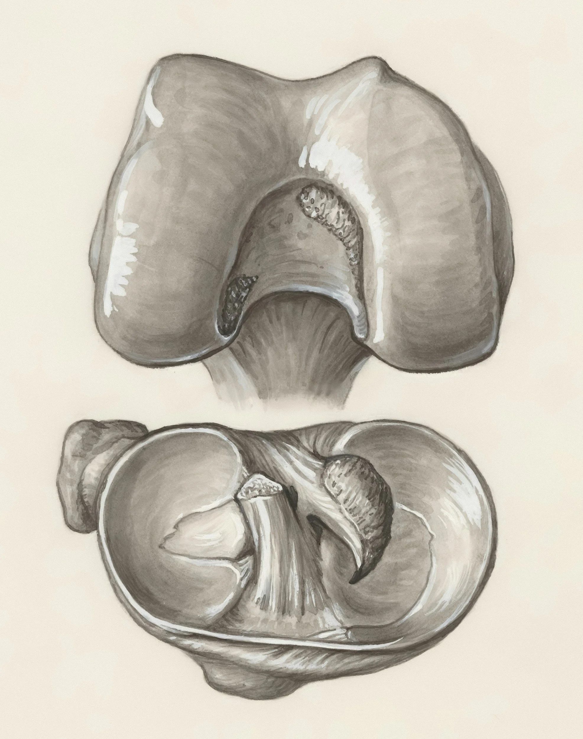
The patellar tendon connects the patella to the tibia and is a critical part of the extensor mechanism that straightens the knee.
Common symptoms:
Sudden pain and a pop
Difficulty or inability to perform a straight-leg raise
Swelling and bruising
A palpable gap below the patella may be present
How it is evaluated:
History and physical exam will typically indicate whether an injury to the patellar tendon is present. X-rays help assess patellar position and associated injury; ultrasound or MRI can confirm the diagnosis but is sometimes not necessary.
If surgery is selected:
Complete acute patellar tendon ruptures require repair. Chronic or failed repairs may require reconstruction or augmentation with autograft or allograft tissue.
Recovery expectations:
Rehabilitation protects the repair from excessive knee flexion and active extensor load early, then progressively restores motion, quadriceps strength and functional loading.

Quadriceps Tendon Rupture
The quadriceps tendon connects the quadriceps muscles to the top of the patella and is a critical part of the extensor mechanism that straightens the knee.
Common symptoms:
Sudden pain and a pop
Difficulty or inability to perform a straight-leg raise
Swelling and bruising
A palpable gap above the patella may be present
How it is evaluated:
History and physical exam will typically indicate whether an injury to the quadriceps tendon is present. X-rays help assess patellar position and associated injuries; ultrasound or MRI can confirm the diagnosis but is sometimes not necessary.
If surgery is selected:
Complete acute quadriceps tendon ruptures require repair. Chronic or failed repairs may require reconstruction or augmentation with autograft or allograft tissue.
Recovery expectations:
Rehabilitation protects the repair from excessive knee flexion and active extensor load early, then progressively restores motion, quadriceps strength and functional loading.

Knee Osteoarthritis
Knee osteoarthritis is a degenerative condition involving cartilage, bone, synovium, and other structures of the knee joint.
Common symptoms:
Activity-related pain and stiffness
Swelling or loss of motion
Pain with stairs, squatting or prolonged walking
Bow-legged or knocked-knee deformity in advanced disease
How it is evaluated:
Weight-bearing radiographs (x-rays) are the principal imaging study. MRI is usually unnecessary when history, examination and X-rays establish the diagnosis and treatment plan.
If surgery is selected:
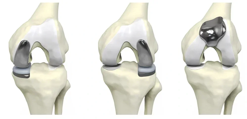
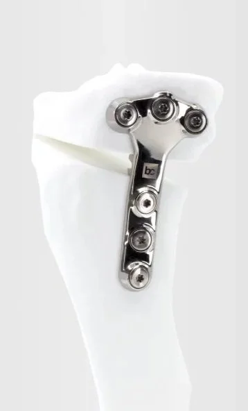
Joint-preserving osteotomy may suit selected younger patients with single compartment overload and malalignment. Unicompartmental or total knee arthroplasty is considered when the disease pattern, pain and functional limitation justify replacement.
Recovery expectations:
After an osteotomy, there is an initial period of protected weight-bearing followed by progressive strengthening and functional rehabilitation. Following partial knee replacement surgery, early weight-bearing is emphasized, with a gradual restoration of motion followed by strength. Traditionally, osteotomy patients are allowed to return to higher impact activities compared to arthroplasty patients, however, the literature in this space continues to evolve.
© Stryker 1998-2026 / https://www.stryker.com/at/en/joint-replacement/systems/mako-partial-knee.html
Bodycad © 2025 / bodycad.com/fine-osteotomy
Patellar Tendinopathy
Patellar tendinopathy is load-related pain and tendon dysfunction, usually at the inferior pole of the patella, common in jumping and running athletes.
Common symptoms:
Localized pain at the lower pole of the patella
Pain with jumping, sprinting, squatting, or stairs
Load-related stiffness or reduced jumping distance
Tenderness at the patellar tendon origin
How it is evaluated:
Diagnosis is usually clinical. Ultrasound or MRI is reserved for atypical presentations, suspected partial tear or refractory symptoms; imaging abnormalities alone do not define symptom severity.
If surgery is selected:
Surgery is uncommon and reserved for persistent, function-limiting symptoms after a prolonged, well-executed loading program and confirmation that the tendon is the primary pain generator.
Recovery expectations:
Recovery after patellar tendon debridement is gradual, with early emphasis on protecting the tendon and restoring knee motion followed by progressive strengthening and tendon loading. Return to running, jumping, and sports is typically staged over several months as pain improves and tendon strength and function recover.
Multi-Ligamentous Knee Injury
A multiligament knee injury (MLKI) is a high-grade injury involving at least two major ligament complexes. It may occur with or without a documented knee dislocation and can be associated with vascular, nerve, meniscus, cartilage or extensor-mechanism injury.
Common symptoms:
Major swelling and instability after trauma
Difficulty bearing weight
Possible numbness or weakness in the foot
A cool/pale foot or abnormal pulses (an emergency)
How it is evaluated:
Initial assessment prioritizes neurovascular status. MRI defines ligament and associated soft-tissue injury; stress radiographs may quantify instability. High-energy injury, bicruciate/PLC patterns, true dislocation or equivocal vascular findings warrant a low threshold for vascular imaging.
If surgery is selected:
Current expert consensus generally favors operative management for many young/active patients with significant MLKI, with repair versus reconstruction, timing and single-stage versus staged surgery tailored to the injury pattern and associated injuries.
Recovery expectations:
Recovery after multiligament knee reconstruction is gradual and highly individualized, with early protection of the repaired or reconstructed ligaments followed by progressive restoration of motion, strength, and stability. Return to running, sports, and higher-level activities typically occurs over many months and depends on the specific ligaments injured, associated procedures, healing, and recovery of strength and function.
MCL Sprain
The medial collateral ligament (MCL) is the primary restraint to valgus stress on the inner side of the knee. Most isolated MCL injuries heal without surgery.
Common symptoms:
Pain and tenderness along the inner knee
Swelling or bruising
Pain with side-to-side stress
Instability in higher-grade injuries
How it is evaluated:
Diagnosis is usually clinical with valgus-stress examination. Radiographs assess for an associated fracture or avulsion. MRI is used selectively, but will certainly be recommended when an ACL or other ligament injury is suspected.
If surgery is selected:
Surgery is uncommon and rarely indicated for an isolated MCL sprain but reconstruction or repair may be considered for select avulsions, Stener-type lesions, persistent valgus/anteromedial rotatory instability or complex multiligament injury.
Recovery expectations:
Recovery after MCL reconstruction is gradual, with early protection of the repaired or reconstructed ligament followed by progressive restoration of motion, strength, and stability. Return to running, sports, and higher-level activities typically occurs over several months and depends on the specific ligaments injured, associated procedures, healing, and recovery of strength and function.